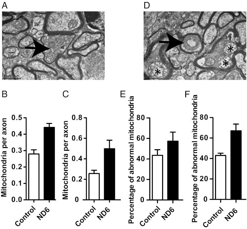
Fig. 2.
Ultrastructural analysis of optic nerve showing mitochondrial abnormalities in ND6 mutant mice. Quantification of mitochondria by transmission electron microscopy (EM) (4,000×). (A) The arrow indicates an axon of 14-mo-old ND6 mutant mouse characterized by mitochondrial proliferation and marked thinning of the myelin sheath. (B and C) Number of mitochondria per axon in optic nerves from 14-mo-old mice [control (n = 4): 267 mitochondria in 954 axons; mutant (n = 4): 427 mitochondria in 964 axons] (B) and 24-mo-old mice [control (n = 3): 188 mitochondria in 723 axons; mutant (n = 4): 384 mitochondria in 801 axons] (C). See Fig. S5A for mitochondrial proliferation at 24 mo. (D) Abnormal mitochondrial morphology as seen with central vacuolization (arrow) and disrupted cristae (asterisks) in 14-mo-old ND6 mutant. See Fig. S5B for mitochondrial abnormalities at 24 mo. (E and F) Percentage of abnormal mitochondria in the optic nerves of 14-mo-old mice (E) and in the optic nerves of 24-mo-old mice (F).

