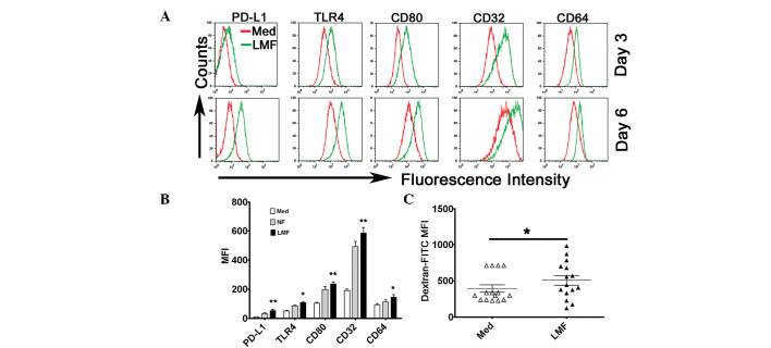
Figure 3.
LMFs regulated PD-L1, TLR4, CD80, CD32 and CD64 expression in monocytes. (A and B) Monocytes were Med or cocultured with NF or LMFs for different time periods. The histograms are representative of 6 separate experiments. (B) Statistical analysis of the MFI with regard to the expression of the surface markers, PD-L1, TLR4, CD80, CD32 and CD64, on the monocytes following 6 days of coculture. (C) The monocytes were left untreated or pretreated for 6 days with the LMFs and were subsequently incubated for 30 min with FITC-dextran at the indicated concentrations (ng/ml). The endocytotic function of the monocytes was assessed by flow cytometry. The values in (B) and (C) represent the mean ± SEM of 6 separate experiments. *P<0.05 and **P< 0.01 indicate significant differences from the untreated monocytes (B and C). LMF, liver myofibroblast; Med, untreated; NF, normal skin fibroblasts.

