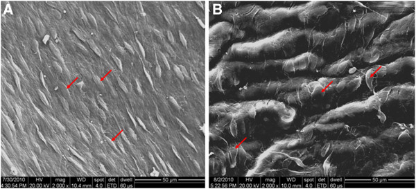
Figure 11.

Endothelial cells on the internal surfaces of grafts harvested 8 weeks after the surgery(× 2000). Red arrows indicate endothelial cells. (A) A PU graft showing complete and continuous endothelial layer lining the inner surface of the graft, well-oriented along the direction of blood flow. (B) A PTFE graft showing complete endothelial layer with disordered arrangement and lack of polarity orientation.
