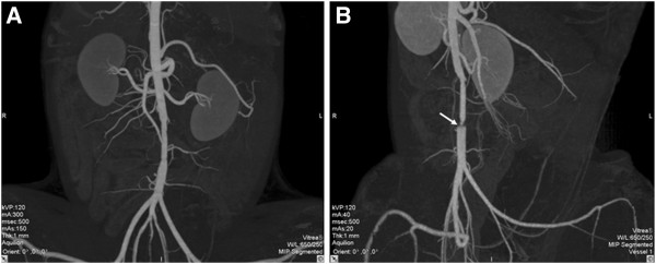
Figure 4.

CT images taken 24 weeks after the surgery(i.e.,shortly before the sample harvest.) (A) A PU graft showing excellent patency, smooth internal wall, and absence of stenosis. (B) A PTFE graft showing patency, yet clear stenosis at the distal anastomotic site (marked with white arrow).
