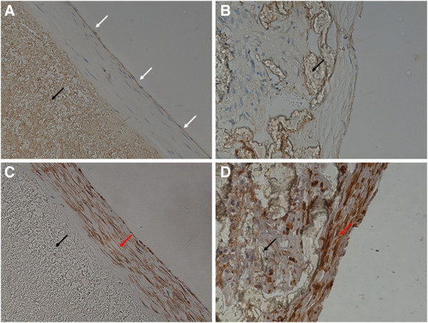
Figure 8.

Neointima on grafts harvested 4 weeks after the surgery(× 400). White arrows indicate endothelial cells, black arrows vascular grafts, and red arrows smooth muscle cells. (A) A PU graft stained for von Willebrand factor (vWF) showing continuous endothelial layer. (B) A PTFE graft stained for vWF showing discontinuous endothelial layer. (C) A PU graft stained for α-SM-actin showing an orderly arrangement of smooth muscle cells. (D) A PTFE graft stained for α-SM-actin showing disordered arrangement of smooth muscle cells.
