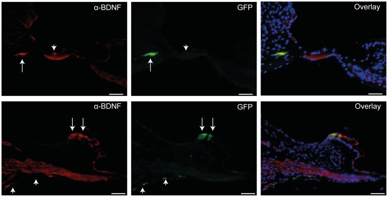
Figure 3. BDNF expression in deafened GPs injected with Ad-GFP-NT at 11 weeks.
Two examples (top and bottom) showing anti-BDNF antibody staining (red) co-localised with GFP expression (green) in some (arrows), but not all cases (arrowheads). As GPs were injected with a combination of vectors encoding for BDNF and NT3, not every GFP-positive cell would be expected to co-localise with BDNF staining, as illustrated in overlays above. Scale bars = 50 µm.

