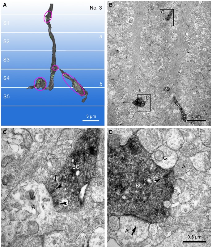
Figure 2. A comparison of axonal synapses and axon terminal synapses of the calbindin ON cone bipolar cell.
A: The 3D reconstruction model for No. 3 calbindin ON cone bipolar cell axon (Fig. 3; Table 1). Magenta ellipses are the labeled profiles shown in B. B: Low magnification view of an electron micrograph used to make the 3D reconstruction model shown in A. Four immunolabeled axonal parts are seen. Among them, one (C) in the upper part of the figure is located in stratum 1 and the remaining three in the lower part are located in strata 4 and 5. The descending axonal part in stratum 1 and an axon terminal part in stratum 5 are depicted and magnified in C and D, respectively. C: The labeled axon descends obliquely to form an axonal ribbon synapse onto an amacrine process (A) represented as a monad. At this synapse, two synaptic ribbons (arrowheads) are engaged. Brackets indicate a putative ribbon in transport. D: A calbindin labeled bipolar axon terminal with a synaptic input from an amacrine cell (A) (arrow) forms a ribbon synapse (arrowhead) onto a postsynaptic dyad composed of a ganglion dendrite (G) and an amacrine process (A).

