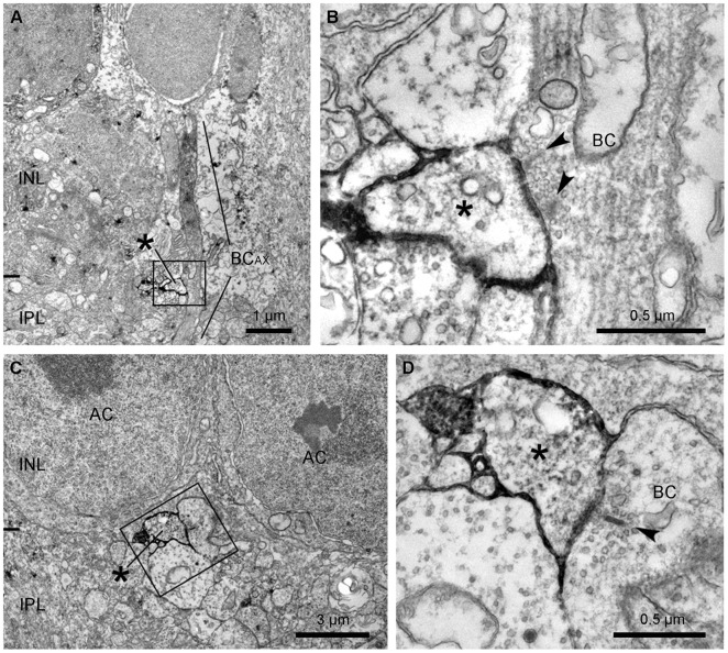
Figure 7. Identification of the synapse between an ipRGC and a putative ON cone bipolar cell in the IPL.
A: A low magnification electron micrograph shows the relationship between a labeled ipRGC dendrite (asterisk) and an unlabeled axon of a putative ON bipolar cell (BCAX), which is running vertically through the INL and sublamina a of the IPL. The boxed area is shown in B at a higher magnification. B: The unlabeled ON bipolar axon (BC) makes an en passant ribbon synapse onto a labeled ipRGC dendrite (asterisk). Note that two synaptic ribbons (arrowheads) are engaged in this monadic synapse. C: A low magnification electron micrograph illustrates a labeled ipRGC dendrite (asterisk) located just below the somata of amacrine cells (AC). The boxed area is shown in D at a higher magnification. D: A labeled dendrite (asterisk) makes a postsynaptic monad at the ribbon synapse (arrowhead) from the axon of a presumed ON bipolar cell (BC).

