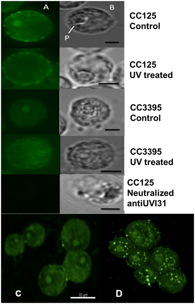
Figure 3. Localisation of UVI31+ protein in C. reinhardtii cells.
Immunofluorescence staining of UVI31+ protein in C. reinhardtii CC125 and CC3395 (from dark phase) and 160 J/m2 UV treated cells (A) and bright field images (B) (Bars 5 µm). 3D rendering of UVI31+ immunofluorescence confocal images of CC3395 control (C) and 160j UV treated cells (D).

