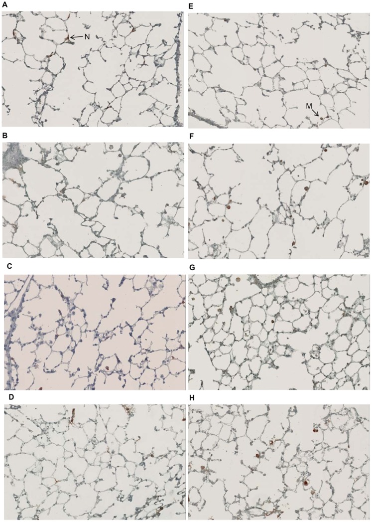
Figure 6. Effect of GPI-0100 on lung histology.
On day 0 and on day 20 mice received either HBS buffer (A, C, E, G) or GPI-0100 at the indicated doses (B, D, F, H) via the intranasal (A, B, E, F) or the intrapulmonary (C, D, G, H) route. Lung samples were collected 3 days after the second treatment upon termination. Lung sections were prepared and stained for Ly-6G and Ly-6C (A-D) or CD68 (E-H) to identify neutrophils and macrophages, respectively. Representative pictures of histological analyses of each treatment group are shown. The brown colored cells indicated by an arrow is a neutrophil (N) or a macrophage (M).

