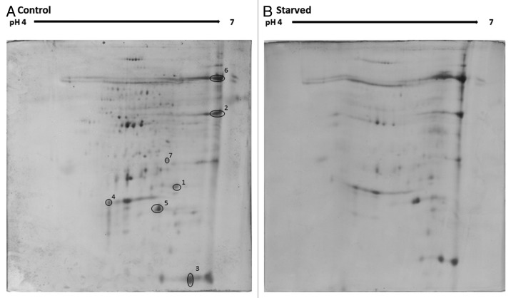
Figure 5. Proteomic profiles from hemolymph of unstarved larvae (A) and larvae starved for 7 d (B). Protein was extracted from larval hemolymph as described and resolved by 2D SDS-PAGE (300 µg/250 µL was loaded onto each Immobiline Drystrip). Peptide spots showing alterations in expression were extracted and identified.

An official website of the United States government
Here's how you know
Official websites use .gov
A
.gov website belongs to an official
government organization in the United States.
Secure .gov websites use HTTPS
A lock (
) or https:// means you've safely
connected to the .gov website. Share sensitive
information only on official, secure websites.
