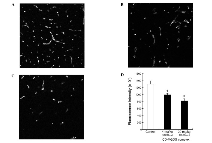
Figure 4.
Immunofluorescence staining for CD31 in Colon26 solid tumor tissue. Groups of mice were treated with (A) CD (200 mg/kg) as a control group (vehicle control, n=6), (B) CD (40 mg/kg)-MGDG (4 mg/kg) complex (n=6) or (C) CD (200 mg/kg)-MGDG (20 mg/kg) complex (n=5). (D) Mean fluorescence intensity values of CD31. *Significantly different from the control, P<0.05. CD, γ-cyclodextrin; MGDG, monogalactosyl diacylglycerol.

