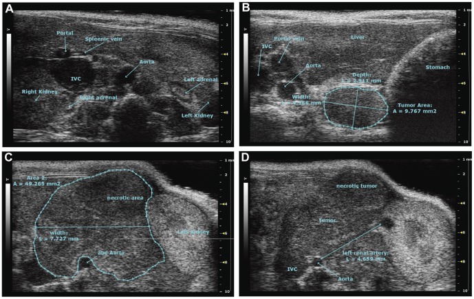
Figure 2. Tumor imaging using high resolution ultrasound.
A. Tumor free animal visualizing abdominal landmarks shown by high resolution ultrasound. B. Detection of a non-palpable small tumor lesion (9.8 mm2) growing on the left side of the spine attached to the muscles. C and D. Imaging of a large palpable tumor (49 mm2). Growing around the aorta and inferior vena cava (IVC), displacing the left kidney. The pictures shown are transverse sections, oriented with the dorsal side down and the ventral up.

