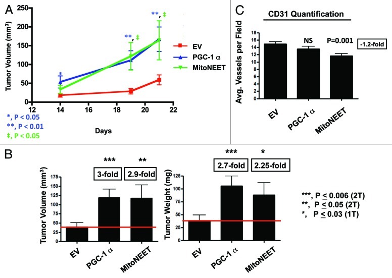
Figure 3. PGC-1α and MitoNEET overexpression increases tumor growth. PGC-1α and MitoNEET overexpressing MDA-MB-231 cells were subcutaneously injected into the flanks of nude mice. (A) Tumor volumes were measured on days 14, 19 and 21. Note that PGC-1α and MitoNEET tumors demonstrate a trend of significantly larger volumes, as compared with control tumors. (B) Tumors were excised, weighed, and their volumes were measured. PGC-1α tumor volumes and weights were 3- and 2.7-fold larger, as compared with control tumors, respectively. Also, MitoNEET tumor volumes and weights were 2.9- and 2.25-fold larger, as compared with control tumors, respectively. (C) Tumor angiogenesis and vessel quantification (number of vessels per field). Frozen sections were cut and immunostained with anti-CD31 antibodies. Quantification of CD31-positive vessels shows a non-significant reduction in angiogenesis in PGC-1α tumors, while MitoNEET tumors displayed a significant reduction in CD31-positive vessels. These results indicate that PGC-1α and MitoNEET stimulate tumor growth, but independently of neo-angiogenesis. EV, empty vector (Lv105).

An official website of the United States government
Here's how you know
Official websites use .gov
A
.gov website belongs to an official
government organization in the United States.
Secure .gov websites use HTTPS
A lock (
) or https:// means you've safely
connected to the .gov website. Share sensitive
information only on official, secure websites.
