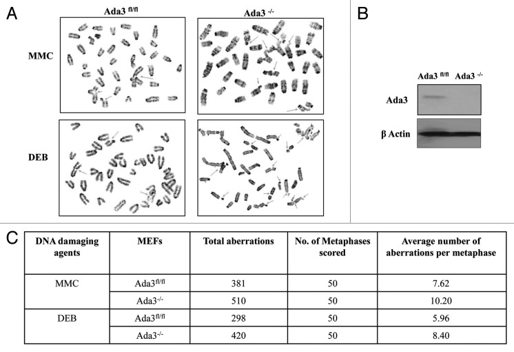
Figure 4. Ada3-null cells are more prone to DNA damage. (A) Representative images depicting chromosome aberrations in Ada3fl/fl and Ada3−/− primary MEFs upon treatment with Mitomycin C (MMC) and Diepoxybutane (DEB). Chromosome aberrations are indicated by arrows. (B) western blotting for Ada3 expression in Ada3fl/fl and Ada3−/− primary MEFs; β-actin was used as the loading control. (C) Summary for MMC- and DEB-induced chromosome aberrations in Ada3fl/fl and Ada3−/− primary MEFs. Giemsa-banded metaphase spreads were analyzed for chromosome abnormalities.

An official website of the United States government
Here's how you know
Official websites use .gov
A
.gov website belongs to an official
government organization in the United States.
Secure .gov websites use HTTPS
A lock (
) or https:// means you've safely
connected to the .gov website. Share sensitive
information only on official, secure websites.
