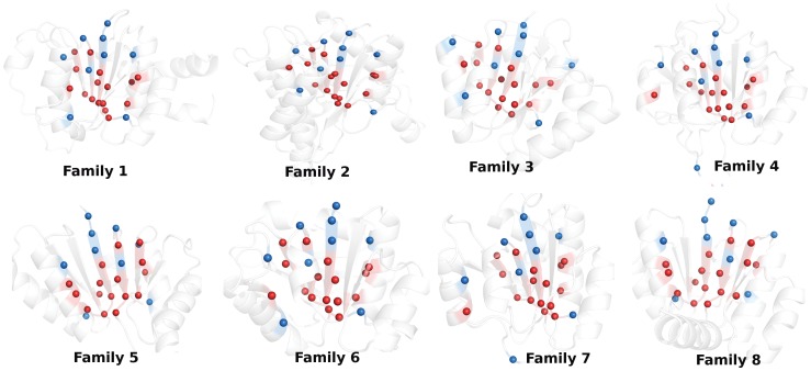
Figure 4. Pictorial depiction of the crucial “fold-specific residues/positions” for one representative member of each of the 8 family under study.
The protein backbone is depicted as new-cartoon and these residues are represented by van der Waals’ spheres. The “fold-specific residues/positions” which are predicted to be important from all the four network parameters (region labeled 1 in Fig. 5) are colored red and those from any combination of three parameters (region labeled 2–5 in Fig. 5) are colored blue. Majority of these residues are either in the solvent exposed termini of the central β-strands or appear along the β-strands.

