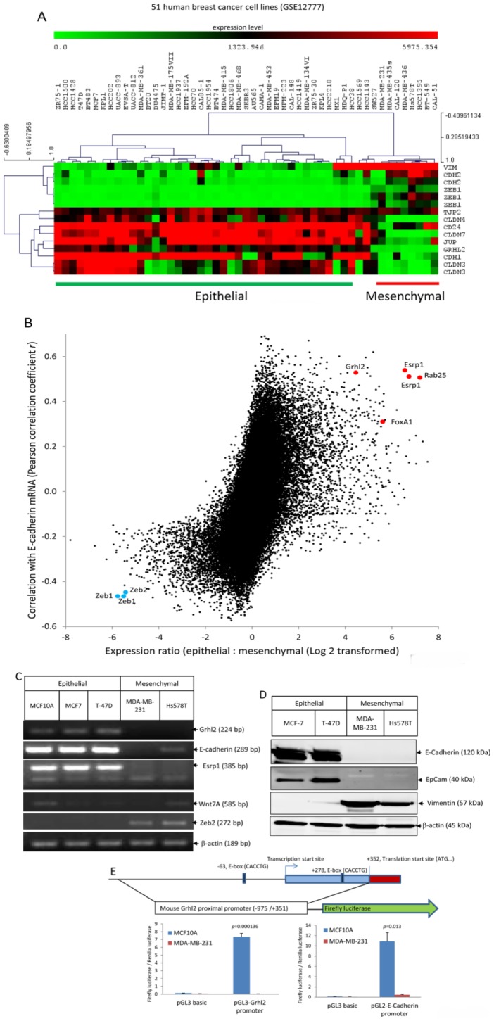
Figure 1. Grhl2 is specifically expressed in epithelial cells and tightly co-regulated with E-cadherin in human breast cancer cells.
(A) According to the molecular definition of epithelial and mesenchymal breast cancer cells [1], [12], [13], the following genes were set as epithelial markers: Cdh1, Cldn3, Cldn4, Cldn7, Tjp2, Jup, and CD24; these genes were set as markers for mesenchymal cells: Cdh2, Vim and Zeb1. Hierarchical clustering (HCL) of 51 human breast cancer cells (Dataset GSE12777) based on transcription levels of epithelial and mesenchymal markers readily separated these cells into two groups: an epithelial group and a mesenchymal group. Grhl2 was a positive significant gene of epithelial cells and a negative significant gene of mesenchymal cells (false discovery rate <5%, two-class significance analysis of microarrays (SAM)). Grhl2 was specifically expressed in epithelial cells with extraordinarily low p-values (p = 1.27558E-07, two tailed student t tests). Red and green squares correspond to high and low mRNA levels, respectively. (B) Microarray based screen for epithelial-specific genes co-regulated with E-cadherin in human breast cancer cells (Dataset GSE12777). All probes representing 54,675 transcripts were analyzed for differential expression between epithelial and mesenchymal cells and Pearson correlation coefficient r was calculated between each transcript and E-cadherin across all microarrays. Some of the transcripts that were highly expressed in epithelial cells and co-regulated with E-cadherin are labeled in Red, and some of the transcripts that were highly expressed in mesenchymal and negatively co-regulated with E-cadherin are labeled in Blue. (C) Examination of Grhl2, E-cadherin, Esrp1, Zeb2 and Wnt7A expression in human mammary epithelial cells and breast cancer cells by RT-PCR. (D) Expression of E-cadherin, Epcam, and vimentin in human breast cancer cell lines was examined by western blotting. (E) Grhl2 promoter activity in epithelial and mesenchymal breast cells. Upper panel: A schematic map of mouse Grhl2 proximal promoter region and luciferase reporter. Bottom panel: Luciferase reporters containing mouse Grhl2 proximal promoter, E-cadherin promoter, or empty vector (pGL3basic) were transiently transfected into MCF10A and MDA-MB-231 cells with a Renilla luciferase vector for normalization. Dual luciferase activities were measured 48 hours post transfection. Data represent one of three independent experiments. Error bars represent mean ± SEM of duplicated experiments. p values were calculated by two tailed student t test.

