Figure 8.
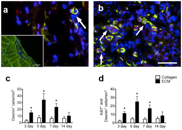
Proliferating muscle cell recruitment. (A) Collagen injection region and (B) skeletal muscle matrix injection region at 5 days with desmin-stained cells (green) co-labeled with Ki67 (red). Arrows denote desmin and Ki67 positive cells. Scale bar at 20 μm. Insert shows positive desmin staining of healthy skeletal muscle, scale bar at 100 μm. (C) Quantification of desmin-positive cells in the skeletal muscle matrix compared to collagen normalized to area. Note that there are significantly more desmin-positive cells in the skeletal muscle matrix. (D) Of these desmin-positive cells, a majority of the cells are proliferating as seen by Ki67 co-labeling.

