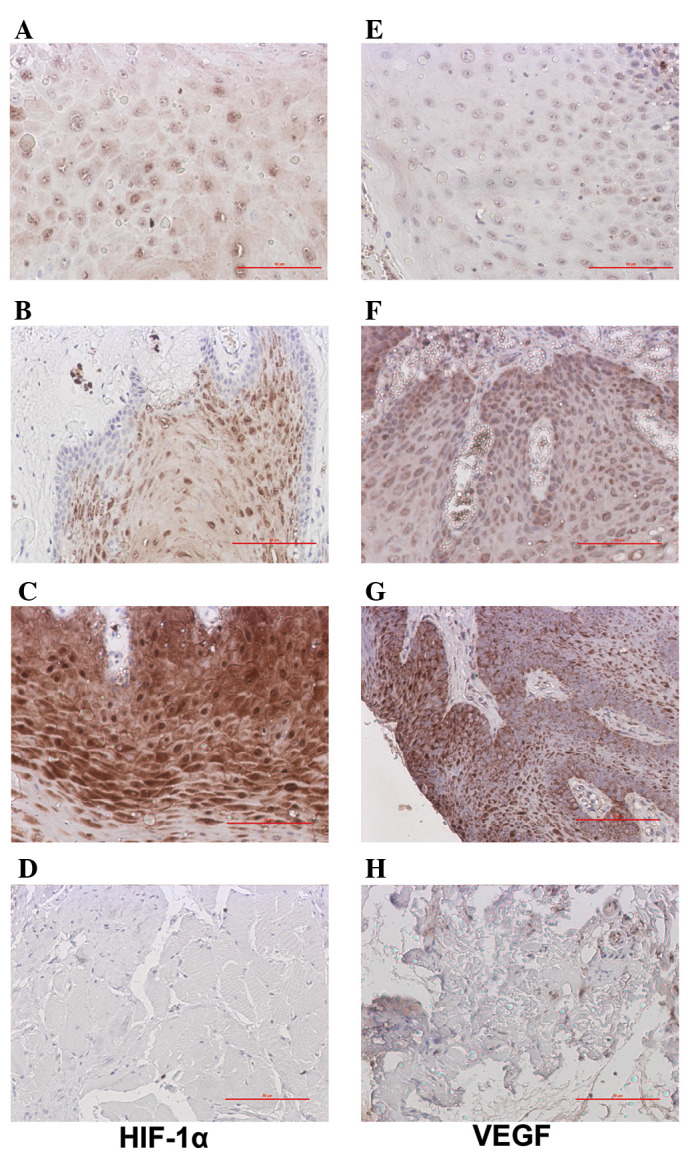
Figure 1.

Expression of HIF-1α and VEGF (immunohistochemisty, magnification, x200). (A–C) Expression of HIF-1α at various levels in TSCC: (A) weak positive, (B) medium positive and (C) strong expression. (D) No expression of HIF-1α in the adjacent tissue. (E–G) Expression of VEGF in TSCC. (H) Weak expression of VEGF in adjacent normal tissue. Red lines, 50 μm; HIF-1α, hypoxia-inducible factor-1α; VEGF, vascular endothelial growth factor; TSCC, tongue squamous cell carcinoma.
