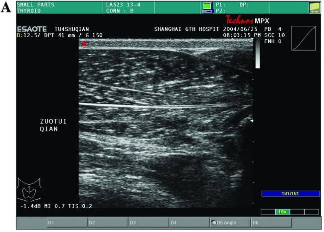
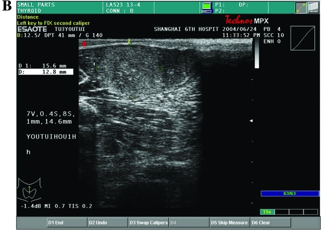
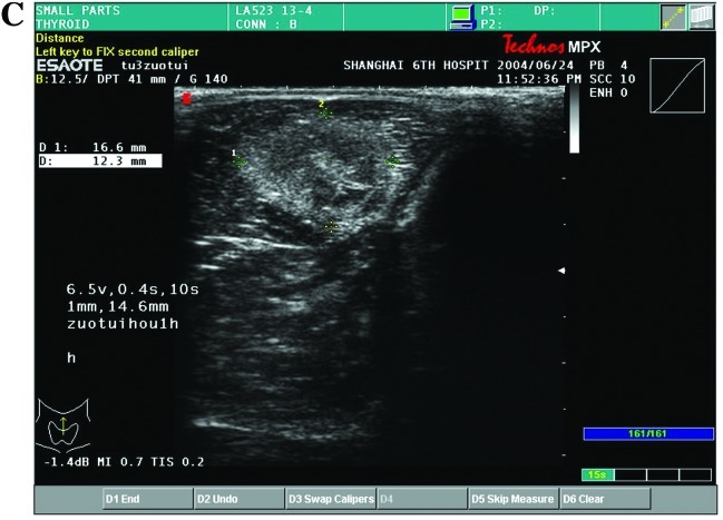
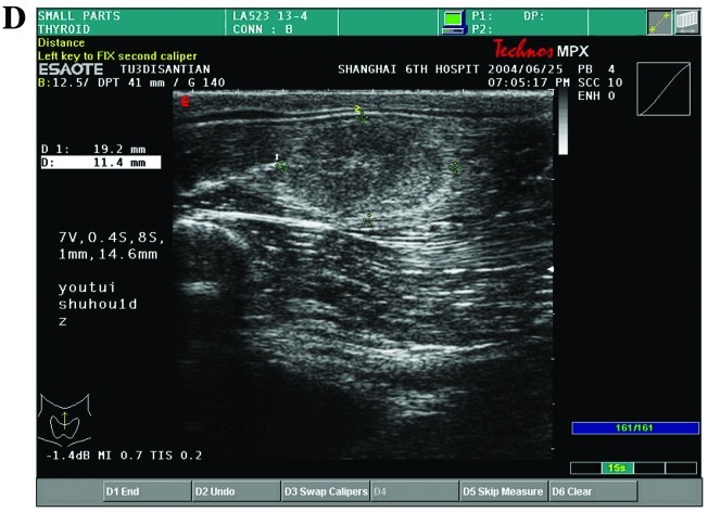
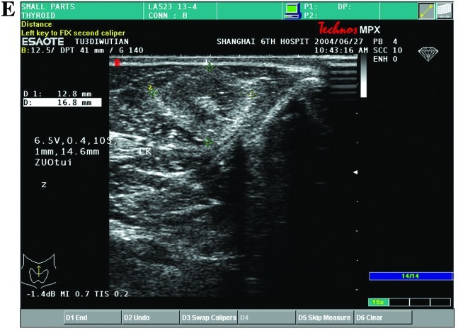
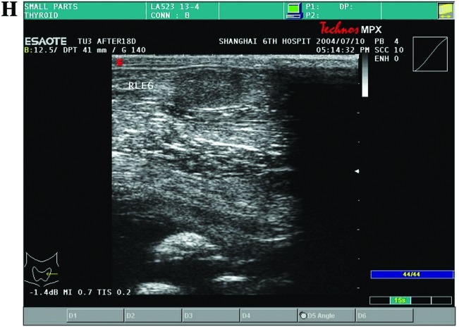
Figure 1.
(A) The echo of rabbit leg muscle was homogeneous and hypoechoic, as detected by the two-dimensional ultrasound prior to HIFU radiation. (B) The echo of the HIFU lesion was hyperechoic at 10 min after HIFU radiation. (C) The echo at the center of the HIFU lesion became lower 1 h after HIFU radiation, there was a distinct hyperechoic border zone around the sonication lesions. (D and E) One to three days later, the echo of sonication lesions became lower. (F) One week later, there was a hypoechoic border zone surrounding the area of sonication. (G and H) Two to four weeks later, the volume of sonication lesions decreased gradually. The echo of sonication-injured tissue was homogeneous and hypoechoic.

