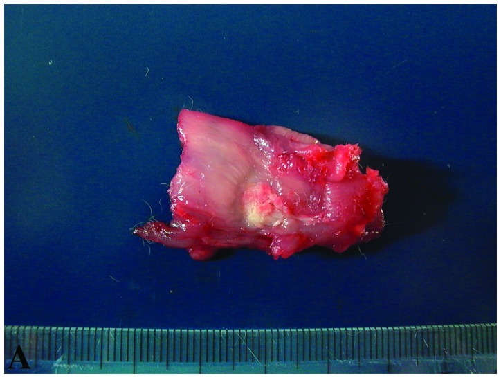
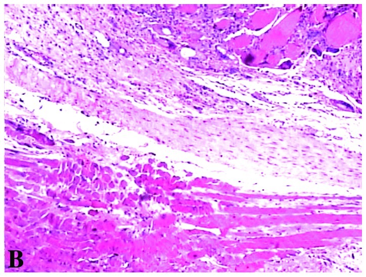
Figure 3.


(A) Four weeks later, a discrete HIFU lesion was observed, with sharp differentiation between normal and ablated tissue. The lesion was best observed grossly as a pale-yellow round area and without hyperemia. (B) The leukocyte and lymphocyte infiltration were not apparent in or around the areas of sonication. There were fibroblast hyperplasia, fatty infiltration and small scar formation in and surrounding the areas of sonication (H&E, ×200 magnification).
