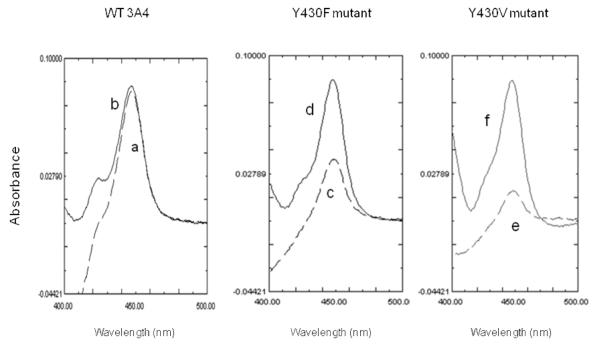
Fig. 9.
Comparison of the P450-CO spectra reduced by NADPH/CPR versus dithionite for WT 3A4 and the Y430F and Y430V mutants. The reduced-CO difference spectra of the three P450s were determined after the samples were reduced by NADPH/CPR and then bubbled with CO (dashed lines a, c and e for WT 3A4, Y430F, and Y430V, respectively). Additional spectra (solid lines b, d, and f for WT 3A4, Y430F, and Y430V, respectively) were obtained following the addition of dithionite as described in Materials and Methods.

