Figure 1.
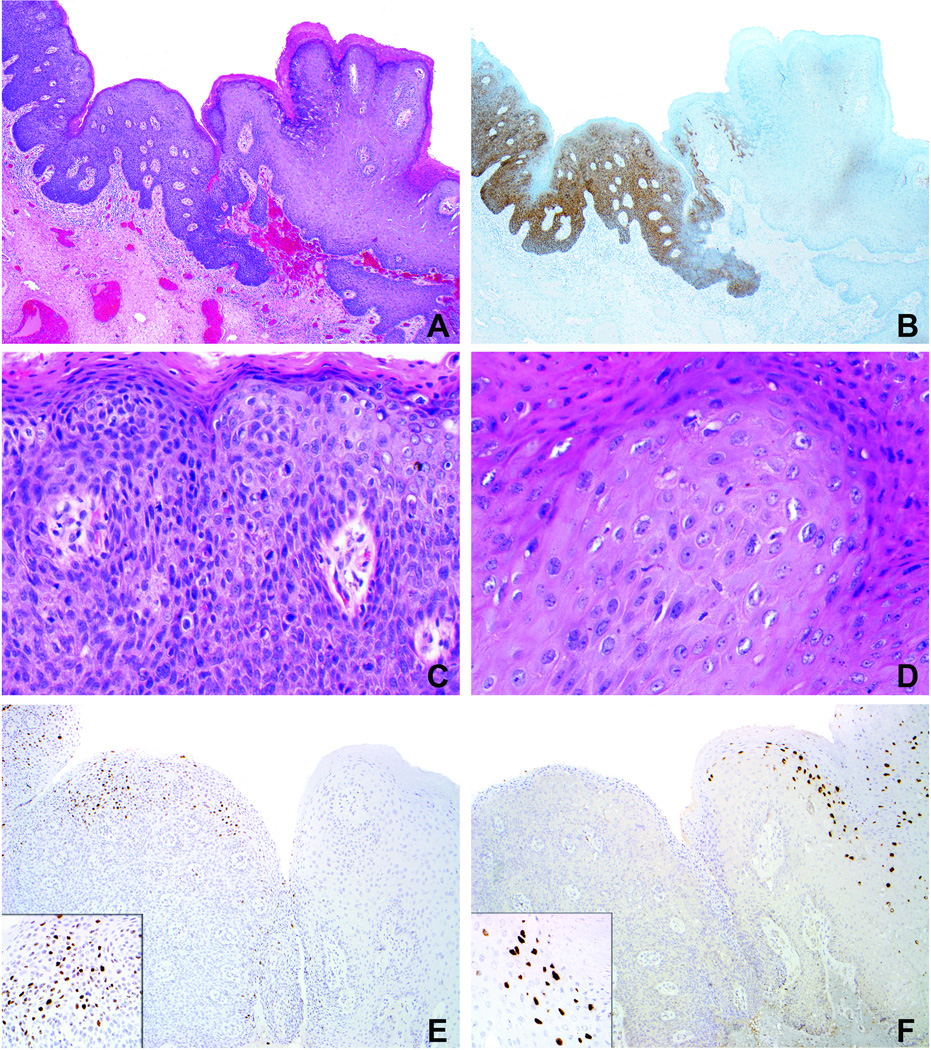
Case 3 (Table 1). A. Condyloma acuminatum (right) with adjacent flat high-grade VIN (left). B. Diffuse p16 expression in high-grade VIN and extremely focal to negative staining in condyloma. C. High-grade VIN exhibits loss of maturation, marked nuclear atypia, and frequent mitotic figures. D. Condyloma is composed of more mature keratinocytes with enlarged nuclei and some koilocytotic change. E. In situ hybridization preparation demonstrates nuclear signals with the HPV 16 probe in the area of high-grade VIN (no signal in condyloma). F. In situ hybridization preparation demonstrates nuclear signals with the HPV 6/11 probe in the area of condyloma (no signal in high-grade VIN).

