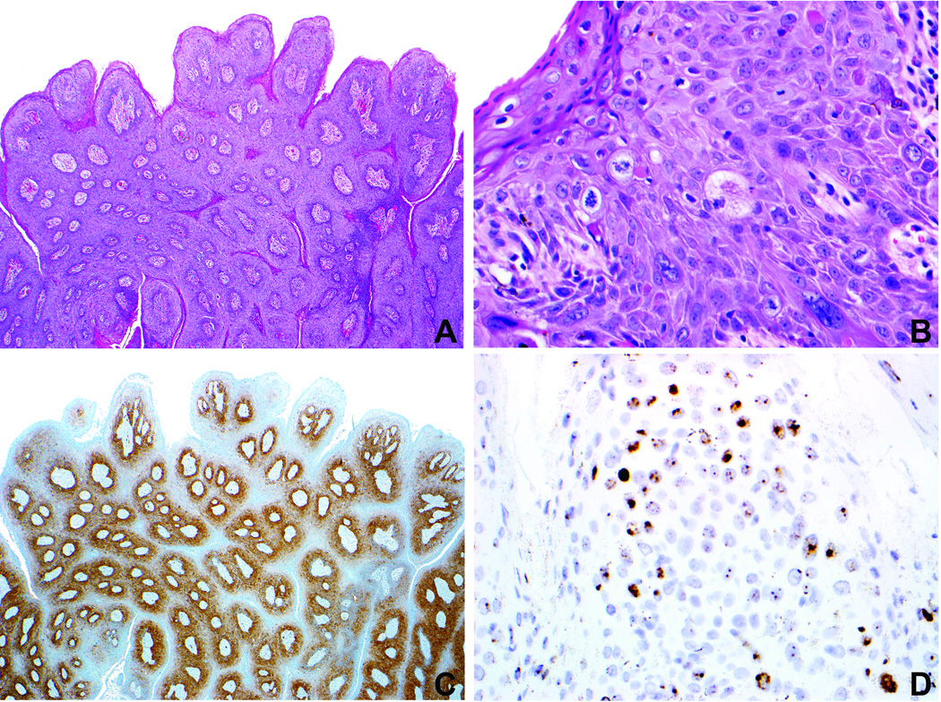
Figure 3.
Case 1 (Table 2). A. Condylomatous high-grade VIN demonstrates striking papillary architecture at low magnification, simulating a condyloma acuminatum. B. Immaturity and notable nuclear atypia consistent with high-grade VIN are evident at higher magnification. C. The lesion demonstrates diffuse p16 expression. D. In situ hybridization preparation demonstrates positive signals with the HPV wide spectrum probe [HPV types 6,11,16,18,31,33,35,45,51,52] and HPV 18 (not shown). There was no detectable HPV with the HPV 6/11 probe (data not shown). These findings support a diagnosis of a high-risk HPV-related high-grade VIN.

