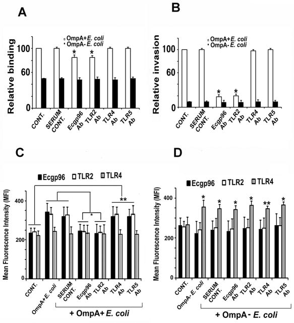
Figure 4. Effect of antibodies to Ecgp6 and TLRs on the binding, invasion and surface expression of Ecgp96.
HBMEC grown in 24 well plates to confluence were pre-incubated with normal serum or antibodies to Ecgp96, TLR2, TLR4 or TLR5 for 1 h and then infected with OmpA+ E. coli K1. Bound (A) or invaded (B) bacteria were determined as described in Methods sections. Data represent mean ± SD from three different experiments performed in triplicate. Relative binding or invasion was expressed with respect to control cell values taken as 100%. The decrease in binding and invasion of E. coli was statistically significant compared with control, *P<0.01 by Student’s t test. In separate experiments, HBMEC were pre-treated with respective antibodies for 1 h prior to infection with OmpA+ E. coli and subjected to flow cytometry analysis using antibodies to Ecgp96, TLR2 or TLR4. Increase or decrease in the expression of the respective proteins in the presence of various antibodies were statistically significant compared to control (*P<0.05 by Student’s t test).

