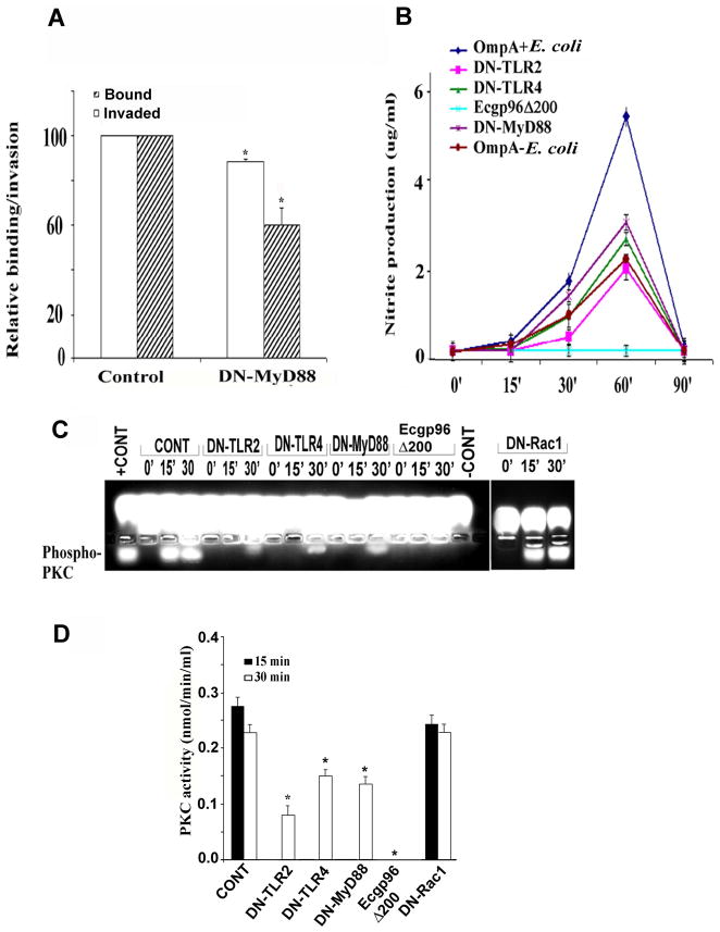
Figure 7. Overexpression of dominant negative-TLR2, -TLR4 or -MyD88 inhibits the production of nitric oxide and activation of PKC-α.
(A) HBMEC transfected with DN-MyD88 or plasmid alone (control) were grown to confluence and used for binding and invasion assays. The data represent means ± SD from three different experiments performed in triplicate. Relative bound/invaded values were expressed with respect to control cell values taken as 100%. The reduction in the cell association and invasion of E. coli in DN-MyD88/HBMEC was statistically significant compared with plasmid-alone transfected cells, *P<0.02 by Student’s t test. (B) HBMEC transfected with various plasmid constructs were infected with OmpA+ E. coli for varying time points, supernatants were collected and analyzed for NO production as mentioned in Experimental Procedures. The data represent mean ± SD from three different experiments performed in triplicate. (C) HBMEC transfected with various plasmids were infected with OmpA+ E. coli for 0, 15 or 30 min, total cell lysates were prepared, and subjected for PKC substrate phosphorylation assay using PepTag non-radioactive kit. +ve represents the positive standard provided by the manufacturer. −ve represents a reaction performed without cell lysates. (D) Spectrophotometric analysis of phosphorylated PKC substrate bands was determined as described in Experimental Procedures. The reduction in the phosphorylation of PKC substrate in DN-TLR2, DN-TLR4, DN-MyD88 and Ecgp96Δ200 transfected and E. coli K1 infected HBMEC was statistically significant, *P<0.05 by Student’s t test.

