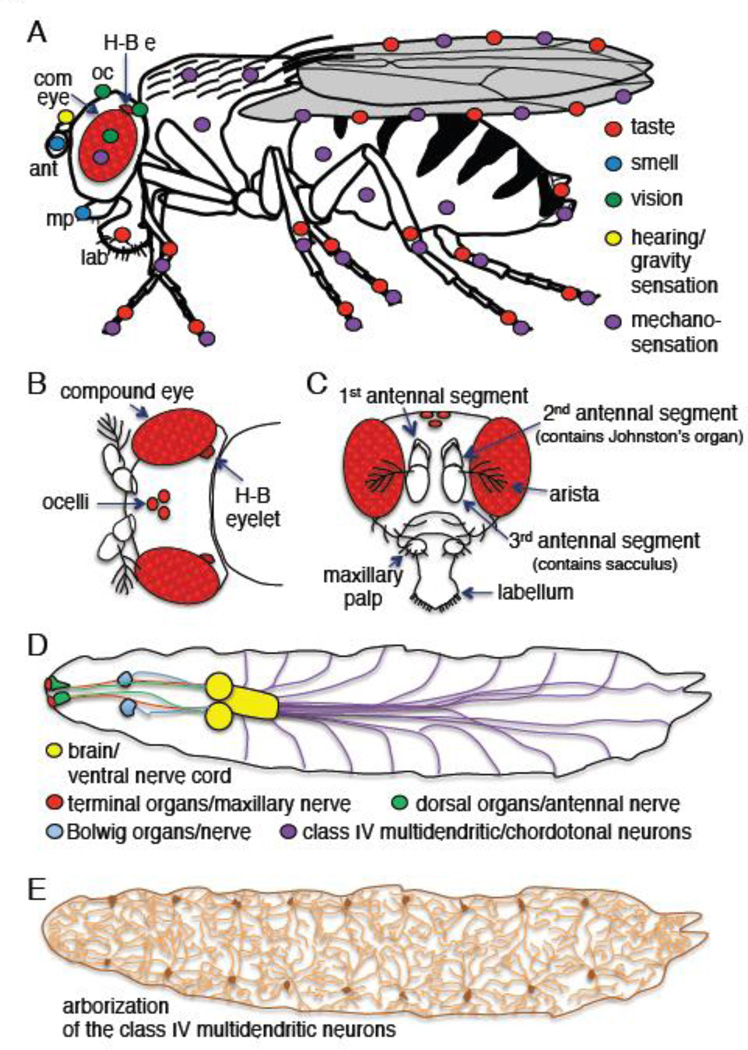
Figure 3. Drosophila sensory organs.
A) Sensory organs in an adult fruit fly. Colored circles indicate the spatial distributions of the sensory organs. Abbreviations: ant, antennae; com. eye, compound eye; H-B e, Hofbauer-Buchner (H-B) eyelet; mp, maxillary palp; lab, labellum; oc, ocelli. The H-B eyelet is located internally in the brain between the retina and lamina, but is shown superficially here and panel B. B) Top view of a fly head showing the visual organs. C) Frontal view of a fly head. D) Larval sensory organs. The cartoon is adapted from previous drawings (Gomez-Marin and Louis, 2012). E) Arborization of the class IV multidendritic neurons that tile the body wall of larvae.

