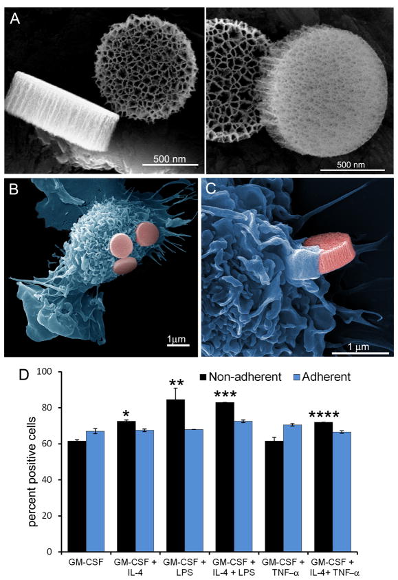
Figure 1. Association of APC with discoidal porous silicon microparticles.
A) Scanning electron micrographs show three perspectives of the 1000 × 400 nm discoidal silicon microparticles, emphasizing microparticle geometry and pore size, with an average pore diameter of 50 nm (left: 160kx mag; 7.5keV; right: 250kx mag; 7.5keV; bars 500 nm). B-C) Pseudo-colored scanning electron micrographs show microparticle association with GM-CSF-stimulated APC at the adherence (B) and initiation (C) stages of uptake (left: 15kx mag; right: 40kx mag; 15 kV; bars 1000 nm). D) Flow cytometry was used to measure association of fluorescent microparticles with both adherent and non-adherent populations of bone marrow-derived APC cultured in the presence of various combinations of GM-CSF, IL-4, TNF-α, and LPS (populations significantly greater than GM-CSF treated cells, *p<0.005, **p<0.04, ***p<0.0006, ****p<0.003). The gamma levels were adjusted on the electron micrographs to enhance contrast and brightness.

