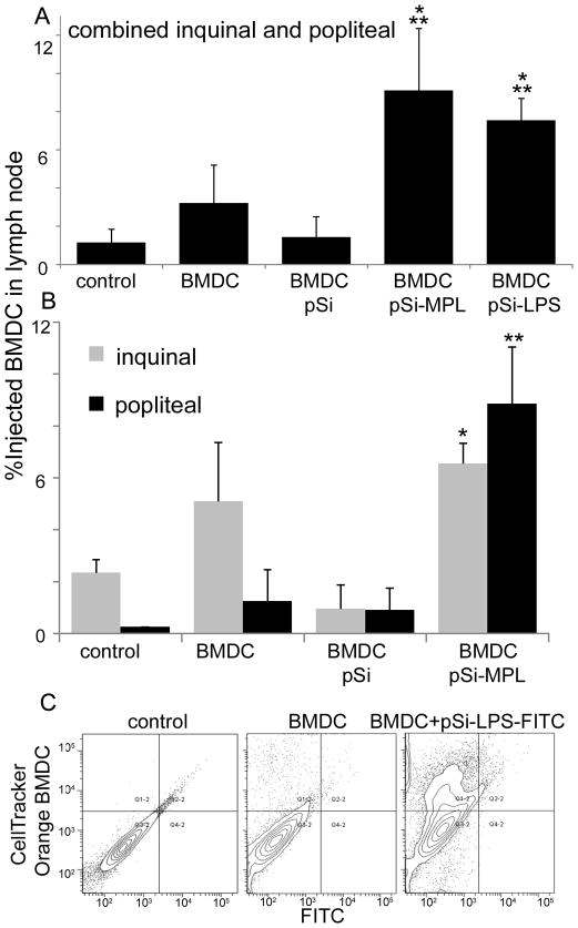
Figure 6. Migration of BMDC to the draining lymph node following subcutaneous injection of microparticle-loaded cells.
A) The percent of combined inguinal and popliteal lymph node cells that are positive for injected (CellTrace™ Green- or PKH26-labeled) GM-CSF-stimulated BMDC 24 hr after injection of mice with 3–5 × 10⁶ control or microparticle-laden BMDC via ipsilateral hock injection (n=5 per group; *p<0.001 compared to BMDC, **p<0.0007 compared to no treatment controls). B) Comparison of microparticle-laden BMDC migration to the popliteal and inguinal lymph nodes following hock injection (*p<0.03, **p<0.04 for BMDC-pSi-MPL compared to non-treated controls). C) Fluorescent contour plots showing inguinal lymph node cell labeling for injected CellTracker™ Orange-labeled BMDC and LPS-FITC bound microparticles (preloaded into the BMDC prior to injection) 24 hr after injection of BALB/c mice with 5 0 × 106 BMDC via hock injection.

