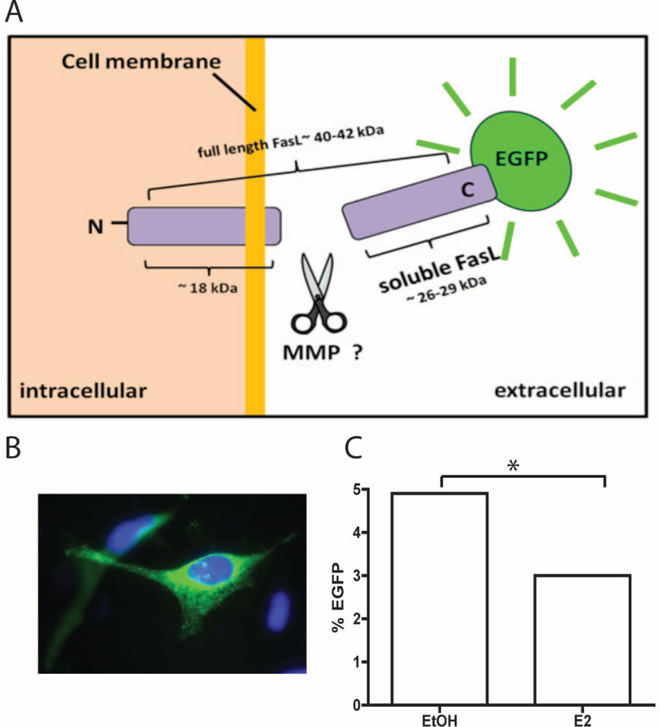
Fig. 1.
EGFP-FasL is cleaved after E2 treatment. (A) Schematic of FasL-EGFP construct. See text for details. (B) U2OS-ERα cells were transiently transfected with FasL-EGFP and then the cells were fixed and stained with DAPI to identify the nucleus. (C) U2OS-ERα cells were transfected with FASL-EGFP then treated with vehicle (EtOH) or 10 nM E2 for 24 hours. Cells were analyzed by flow cytometry and the percent of EGFP-positive cells is graphed. * = p-value <0.001.

