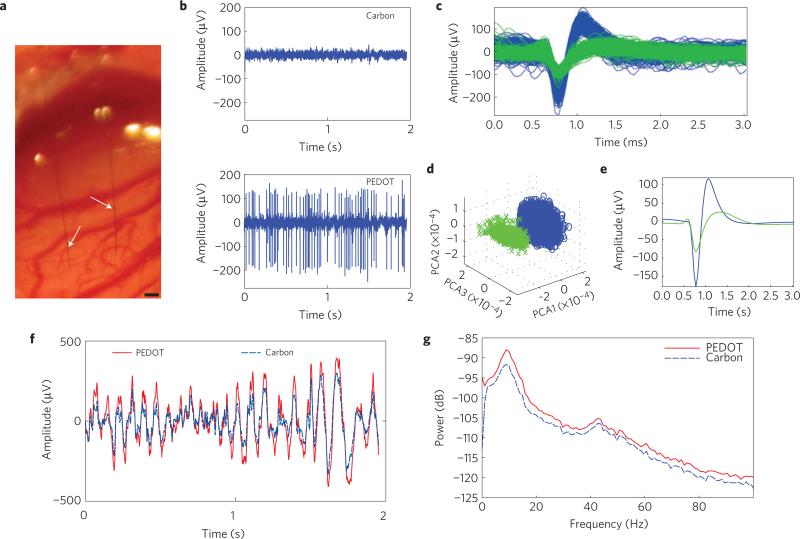
Figure 4. In vivo single-unit recording capabilities.
a, MTA stereotrode (arrows) implanted 1.6 mm deep into the cortex. Scale bar, 100 μm. b, Representative example of 2 s of high-speed recordings taken simultaneously on the same array. Recordings on the carbon site channel show no discernible single units with a noise floor of 16.6 μV (top), whereas recordings taken on the channel with the PEDOT:PSS site show SNRs of 12.7 and 4.71 and a noise floor of 23.4 μV (bottom). c, Piled single-unit neural recordings over 3 min from a poly(p-xylylene)-coated PEDOT:PSS device. d, Results from PCA showing two distinct clusters. e, Mean waveform for each unit spike. f, Raw LFP simultaneously recorded across both channels. The PEDOT:PSS site channel recorded a noise floor of 332 μV (solid red), whereas the carbon site channel recorded a noise floor of 233 μV (dashed blue). g, Power density spectra across the LFP range showing that for the LFP range both recording materials are similar.

