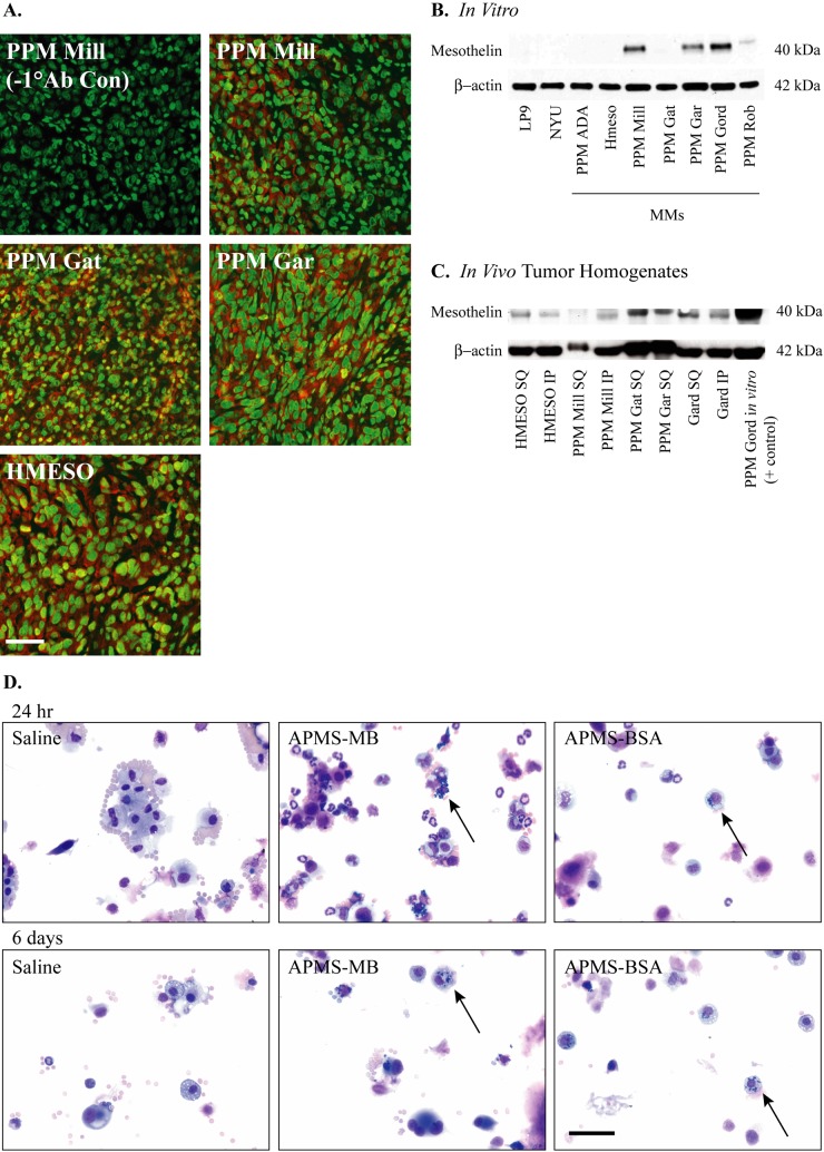
Figure 4.
Mesothelin is expressed in mesothelioma (MM) tumors. Paraffin sections of MM tumors derived from four different MM cell lines (PPM Mill, PPM Gat, PPM Gar, and HMESO) grown in severe combined immunodeficient (SCID) mice were stained with mouse anti-mesothelin antibody (clone MB) (A). Alexa Fluor 568 secondary antibody was used to visualize mesothelin (red), and SYTOX Green nucleic acid stain was used to visualize cell nuclei (green). Micrographs were taken at 400× magnification, scale bar = 50 µm. Mesothelin protein was assessed by Western blot analysis on human MM cell lines (B) and in human MM tumors grown either intraperitoneally (IP) or subcutaneously (SQ) (C). β-Actin was used as a loading control. Micrographs of Hema 3 differential stained cytospins from peritoneal lavage fluid (PLF) show uptake of acid-prepared mesoporous spheres (APMS)–MB and APMS-BSA microparticles (n=5 mice/group). Particles surround the cell nuclei at 24 hr and 6 days (black arrows) (D). Micrographs were taken at 400× magnification, scale bar = 50 µm.

