Figure 6.
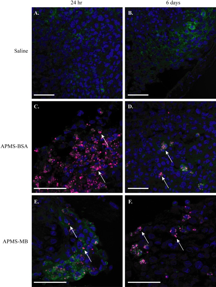
Acid-prepared mesoporous spheres (APMS) microparticles were taken up by mesenteric mesotheliomas (MMs), retained for up to 6 days, and possibly trafficked by tumor-associated macrophages (TAMs) (n=5 mice/group). Confocal microscopy analysis of frozen sections of MM tumor tissue at 24 hr and 6 days shows no particles in saline-injected control tumors (scale bar = 50 µm) (A, B). APMS-BSA and APMS-MB were taken up by tumor tissue at 24 hr and remained in the tumor tissue at 6 days following intraperitoneal (IP) injection (C–F) (scale bar = 50 µm). In all panels, APMS-MB or APMS-BSA are indicated with A647 (red) (white arrows), cell nuclei are stained with DAPI (blue), and TAMs are stained with Mac-2 (green). Panels B, C, E and F are of the tumor exterior (tumor edge) vs. panels A and D of the tumor interior.

