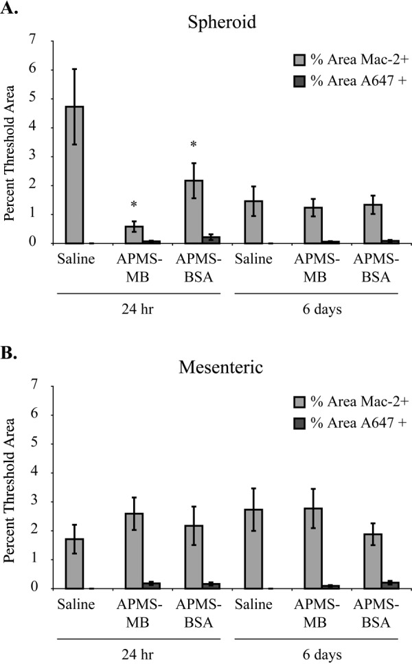
Figure 7.

The percentage of Mac-2+ area within regions of interest analyzed in spheroid mesothelioma (MM) tumor tissue was significantly decreased in acid-prepared mesoporous spheres (APMS)–MB and APMS-BSA treated animals at 24 hr as compared with saline controls (*p<0.05) (n=1 tumor per mouse/3 mice per group) (A). Five randomly chosen regions of interest from tiled (4 × 4 tiles) confocal fluorescent microscopy images were opened in MetaMorph image analysis software to determine differences in the percent of threshold areas positive for the minimum fluorescence intensity of Mac-2 and A647 (APMS preparations). Minimum intensity thresholds were set by Mac-2+ control samples of mouse aorta and visual assessment of multiple A647+ areas. No significant differences were seen in Mac-2+ percent threshold areas by particles in mesenteric MM tissues (B).
