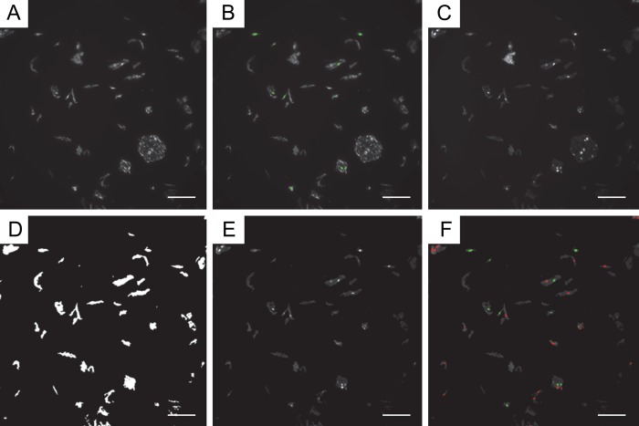
Figure 2.
Representative example of quantification of Acr-dG immunofluorescence using MetaMorph. For immunofluorescence analysis of Acr-dG, formalin-fixed paraffin-embedded (FFPE) oral cells were subjected to a standard procedure using a monoclonal antibody against Acr-dG. Cy3 was used as the secondary antibody, and nuclei were counterstained with 4′,6-diamidino-2-phenylindole (DAPI). Samples were imaged using a HistoRx PM-2000 imaging device, and images were analyzed to identify and enumerate positively stained nuclei using MetaMorph software. (A) Original image in Cy3 channel staining for Acr-dG. (B) Cy3 image; green nuclei are those identified “positive for Cy3” by MetaMorph. (C) Original image in DAPI channel. (D) DAPI image-integrated morphometry mask to eliminate large and small objects. (E) Resulting filtered DAPI image. (F) Final DAPI image; green nuclei are those identified “positive for Cy3” by MetaMorph, and red nuclei are those identified “negative for Cy3” by MetaMorph. The scale bar size shown is 100 µm.

