Figure 6.
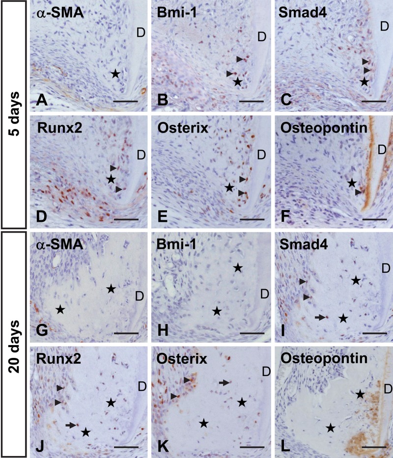
Immunohistochemical localization of α-smooth muscle actin (α-SMA) (A, G), Bmi-1 (B, H), Smad4 (C, I), Runx2 (D, J), Osterix (E, K), and osteopontin (F, L) in the area of root apex at 5 (A–F) and 20 (G–L) days after transplantation. (A) α-SMA-positive cells are not detected at the root apex. (B–E) Some cells (arrowheads) within and around the newly formed apical bone-like tissue (stars) are immunopositive for Bmi-1, Smad4, Runx2, and Osterix. (F) Osteopontin is localized in cells and matrix near the dentin (D). (G, H) Cells around the apical bone-like tissue hardly show immunoreactivity for α-SMA or Bmi-1. (I–K) Cells positive for Smad4, Runx2, and Osterix include lining cells (arrowheads) and cells (arrows) embedded in the apical bone-like tissue (stars). (L) The matrix of the apical bone-like tissue near the dentin demonstrates immunoreactivity for osteopontin. Bar = 50 µm.

