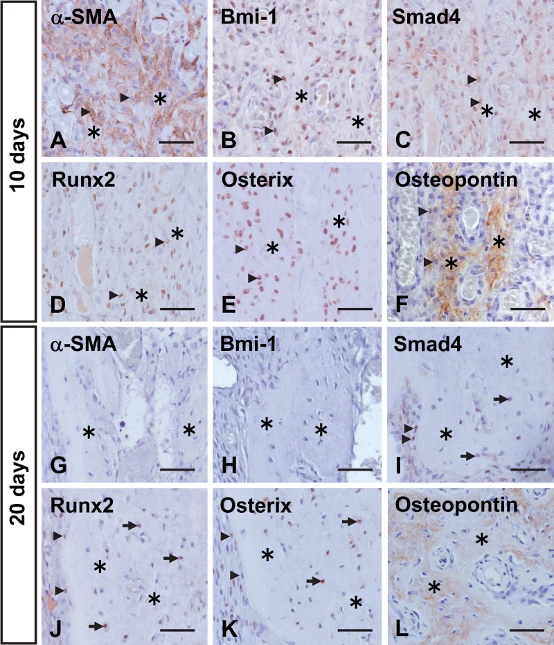
Figure 7.
Immunohistochemical localization of α-smooth muscle actin (α-SMA) (A, G), Bmi-1 (B, H), Smad4 (C, I), Runx2 (D, J), Osterix (E, K), and osteopontin (F, L) in the upper-pulp area at 10 (A–F) and 20 (G–L) days after transplantation. (A) Numerous α-SMA-positive cells (arrowheads) are visible in the upper-pulp cavity. (B–E) Intense staining for Bmi-1, Smad4, Runx2, and Osterix is seen in lining cells (arrowheads) and cells (arrows) embedded in the root bone-like tissue (asterisks). (F) Osteopontin is localized to bone-like tissue matrix and its lining cells. (G, H) There is no expression of α-SMA or Bmi-1 in the upper pulp, except for α-SMA immunoreactivity in its vascular endothelial cells. (I–K) Smad4, Runx2, and Osterix demonstrate a similar pattern of immunoreactivity as that at 10 days after transplantation; however, weaker staining of these cells is observed. (L) The matrix of the root bone-like tissue is immunopositive for osteopontin. Bar = 50 µm.

