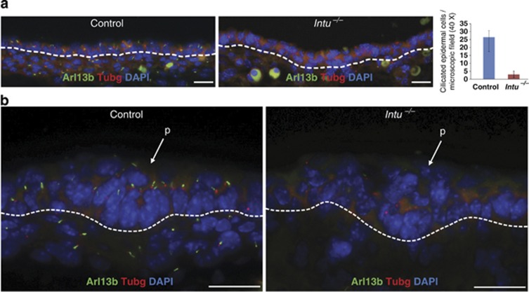
Figure 4.
Primary cilia in dorsal skin of E14.5 Intu−/− mutants. (a) Primary cilia were detected by Arl13b (green) in the epidermal keratinocytes and dermal fibroblasts of Control skins (26.4±6.6 ciliated keratinocytes per microscopic field in Control; 3.0±2.1 in Intu−/− mutants); n=4, P=4.8960 × 10⁻⁸. (b) Keratinocytes of the hair placode (p) were ciliated in Control skin but not in Intu−/− skin. Basal bodies (red) were labeled with γ-tubulin (Tubg); nuclei were stained by DAPI (blue). Dotted line indicates the basement membrane. (a and b) Scale bar, 20 μm

