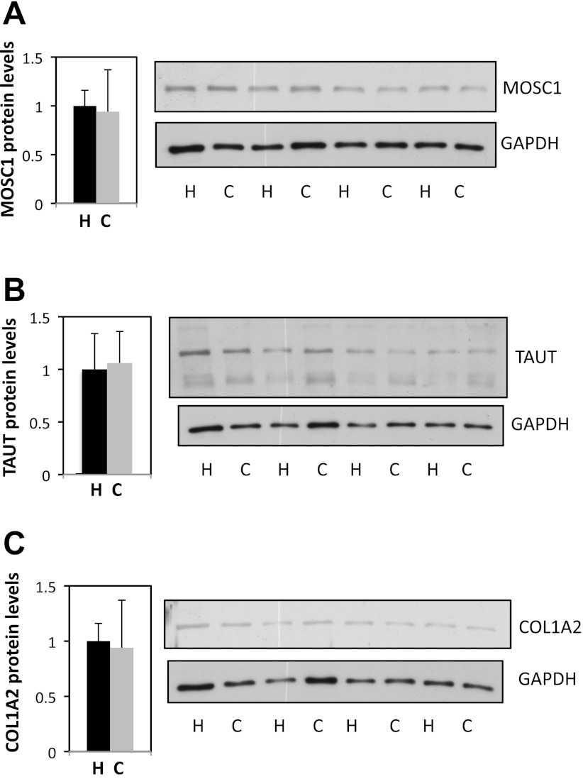
Fig. 4.
Protein levels of MOSC1, TAUT, and COL1A2 in ventricular biopsies of controlled reoxygenation vs. hyperoxic patients. Tissues were lysed to isolate protein content and Western blotting analysis performed probing for MOSC1 (A), TAUT (B), COL1A2 (C), and GAPDH. No significant changes were observed in controlled reoxygenation (C) compared with hyperoxic (H) samples. MOSC1, CRIP3, and COL1A2 bands were normalized to GAPDH levels. Data are means ± SE.

