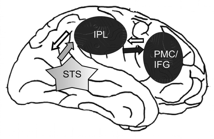
Figure 1.
Neuronal basis of imitation (after [11], modified). The Figure shows the frontoparietal mirror neuron system (MNS) (black ovals) and visual input (grey star) in the human brain. The anterior area of the MNS involves the posterior inferior frontal gyrus (IFG) and the ventral premotor cortex (PMC), and the rostral area involves the inferior parietal lobule (IPL). The grey arrow indicates input to the MNS from the STS. The black arrow shows the information flow from the IPL to the PMC/IFG. The white arrows show the information flow from PMC/IFG to the IPL and to the STS (based on [11]).

