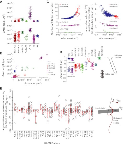
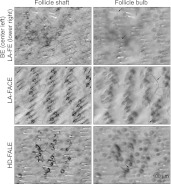
Figure 8. Parametric analysis of arbor area, arbor depth, axon length, and number of follicles innervated across arbor classes.
(A) Arbor areas plotted on linear (top) and log10 (bottom) scales for the seven arbor types with arbor areas larger than the surround of a single follicle (i.e. arbor types other than I-FALE, I-FACE, and MCA). (B) Axon length vs arbor area. Note the break in the horizontal and vertical axes and the compressions in scale beyond the break. (C) Comparisons among the four arbor classes that innervate multiple follicles (HD-FALE, LD-FALE, LA-FACE, SA-FACE): number of follicles innervated vs arbor area, and innervation index (defined as number of innervated follicles per arbor area) vs arbor area. (D) Arbor depth within the skin. The skin surface corresponds to 0 μm. On the back skin at P21, the mean depth of a follicle bulb is ∼100 μm. (E) Orientations of follicle-associated C-shaped lanceolate endings from 34 LD-FALE arbors relative to the orientation of their associated follicle. In wild type mice at P21, hair follicles on the back are oriented from anterior to posterior, with mean deviations from that axis of less than 10 degrees (Wang et al., 2010). Red bars show mean and standard deviation. VNE, vector orientation for the nerve ending; VHF, vector orientation for the hair follicle.


