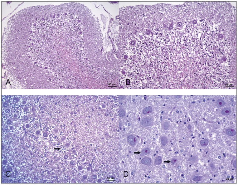
Figure 4.
Photomicrographs of the cerebellum of the dog in case 2. (A–C) Severe depletion of granular neurons with mild decrease in Purkinje cell number, empty baskets, and Bermann’s glia proliferation (arrow) H&E. (D) Presence of PAS-positive material in the perikarya of vestibular nucleus neurons (arrow). PAS.

