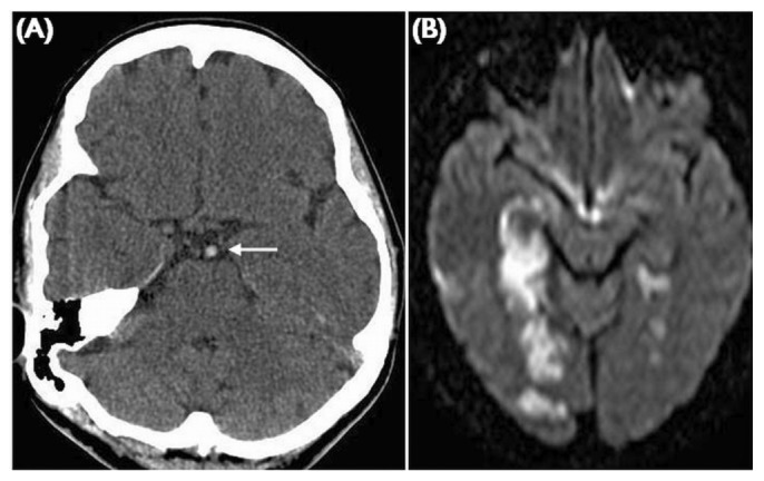
Figure 4.

— Basilar artery thrombosis. (A) Unenhanced computed tomography demonstrates a hyperdense basilar artery indicating acute thrombus (white arrow). The diffusion weighted imaging shows bilateral posterior circulation infarcts involving the white and grey matter.
