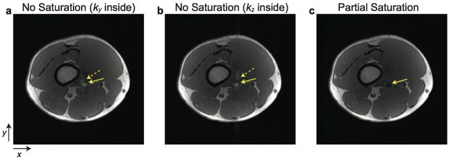
Figure 8.
In vivo leg images. The femoral artery of the leg is depicted by solid arrows. a–b: Without partial saturation. (a) is acquired with ky as the inner acquisition loop, while (b) is acquired with kz as the inner loop. Pulsatile ghost artifacts (dashed arrows) are generated along the outer phase-encode direction. c: With partial saturation, inflow enhancement within the femoral artery and ghost artifacts are greatly reduced.

