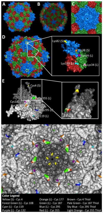
Figure 1. The structure of CPMV.
CPMV consists of 60 copies of a small subunit (S, blue) and a large two-domain subunit (L, green and red). A) Exterior view and B) interior view. C) Pore structure at the five-fold axis; the pore diameter was measured to be 0.75 nm. D) CPMV displays 300 reactive lysines on the surface, five per asymmetric unit. E) CPMV also displays cysteines selectively on the interior surface, 8 per asymmetric unit (Cys 4 – yellow, 108 – forest green, 119 – cyan, 132 – purple, 177 – orange, 187 – green, 295 – blue, and 355 – red). Inset shows 90 degree rotation, revealing hidden reactive thiol of Cys 4. F) View of interior with surface-exposed thiols highlighted (Cys 4 – brown, 187 – pale green, 295 – sky blue, and 355 – light orange).

