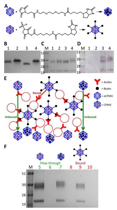
Figure 3.
A) Schematic of biotin functionalization of eCPMV-bioI and CPMV-bioE, respectively B) Biotinylated particles on a 1.2% agarose gel visualized after Coomassie staining. 1=CPMV; 2=eCPMV; 3=CPMV-biotin; 4=eCPMV-biotin. C) Same particles on a 4-12% denaturing SDS-PAGE gel stained with Coomassie. M=SeeBlue Plus2 molecular weight marker. D) Western blot probed with streptavidin-alkaline phosphatase confirms biotinylation. E) Schematic of the avidin bead binding assay. F) Flow through and eluted particles from binding assay on a SDS-PAGE gel after staining with Coomassie. 5=CPMV flow through; 6=CPMV-bioE flow through; 7= eCPMV-bioI flow through; 8=bound CPMV; 9=bound CPMV-bioE; 10=bound eCPMV-bioI.

