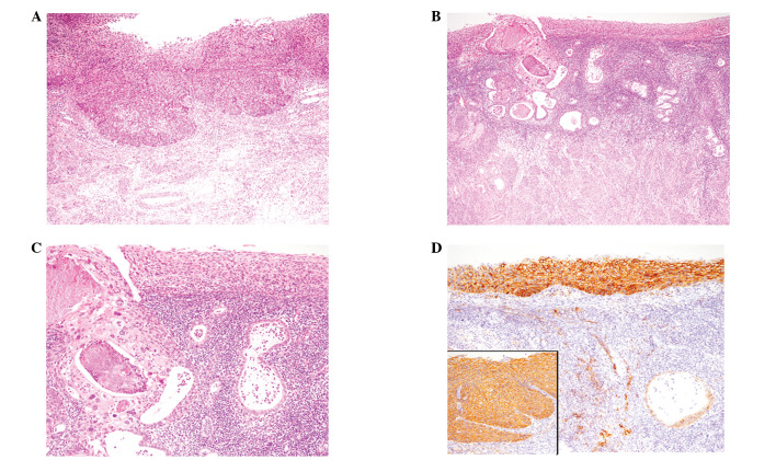
Figure 1.
Histopathological and immunohistochemical features of case 1. (A) Mild invasive growth of atypical squamous cells in the cervix (hematoxylin and eosin, ×100). (B) Low-power view of superficial spreading atypical squamous cells in the endometrium (hematoxylin and eosin, ×100). (C) High-power view showing superficial spreading and glandular involvement of atypical squamous cells (hematoxylin and eosin, ×200). (D) CD138 was strongly expressed in the neoplastic squamous cells spreading in the endometrium (×200). Mildly invasive squamous cells in the cervix also expressed CD138 (inset, ×100).

