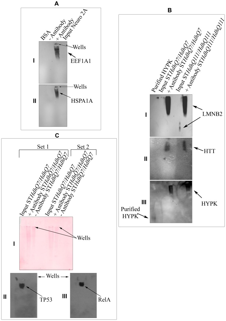
Figure 3. Formation of high molecular weight HYPK-complex.
High molecular weight HYPK-complex formation by endogenous HYPK with EEF1A1 (AI) and HSPA1A/HSP70 (AII) in Neuro2A cells using native-PAGE western analysis; results obtained by similar experiments in STHdhQ7/HdhQ7 and STHdhQ111/HdhQ111 with LMNB2 (BI) and HTT (BII); result obtained by re-probing the blot shown in B with anti-HYPK antibody is shown in BIII, showing the difference in migration of purified HYPK and complex-bound HYPK; similar co-IP experiments in STHdhQ7/HdhQ7 (C) along with Ponceau staining of the nitrocellulose membrane (CI). The blot is probed with anti-TP53 (CII) and anti-RELA (CIII) antibodies. In all the cases, anti-HYPK antibody was used to pull-down and the immunoprecipitate elute was analyzed to detect the interacting proteins. The loading wells in A and C are marked by arrows.

