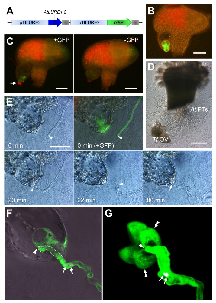
Figure 7. Interspecific pollen tube attraction assay using transgenic T. fournieri ovules and A. thaliana pollen tubes.
(A) Schematic of the sequences introduced into T. fournieri. The TfLURE2 promoter (pTfLURE2) was used to express both AtLURE1.2 and GFP genes in synergid cells. The sequence of the AtLURE1.2 gene is a genomic sequence from the translation initiation codon to the putative 3′ untranslated region. Gray boxes indicate nopaline synthase terminators. (B) GFP expression in two synergid cells of the transgenic T. fournieri ovule. Scale bar, 50 µm. (C) Immunostaining of transgenic T. fournieri ovules with anti-AtLURE1.2 (CRP810_1.2) antibodies. Red Alexa Fluor fluorescence for AtLURE1.2 (arrow) was observed at the micropylar surface of the embryo sac of the ovule labeled with GFP (left panel). In the ovule without GFP labeling (right panel), a weaker pseudo-signal was observed at the micropylar surface of the embryo sac. Scale bars, 50 µm. (D) An overview of the pollen tube attraction assay. Pollen tubes of A. thaliana (At PTs) are observed emerging from a cut style and growing across the medium. A manipulated T. fournieri ovule (Tf OV) was placed near the out-growing pollen tubes. Scale bar, 200 µm. (E) Pollen tube attraction toward the synergid cell of a transgenic T. fournieri ovule. Arrowheads indicate the position of the tip of the A. thaliana pollen tube. At 0 min, an ovule was placed in front of the tip of the pollen tube using a glass needle. The ovule expressed GFP in the synergid cell, indicating co-expression of the AtLURE1.2 peptide. Green fluorescence in the pollen tube indicates pollen-expressed pLAT52::GFP. The pollen tube was attracted to the ovule (20 min). After shifting the position of the transgenic ovule (22 min), the pollen tube redirected itself once again toward the micropyle (80 min). The arrow indicates the ovular micropyle penetrated by the attracted pollen tube. Scale bar, 50 µm. (F) A confocal laser scanning microscopic (CLSM) image of the transgenic T. fournieri ovule shown in (E), demonstrating that the tip of the A. thaliana pollen tube has fully penetrated the transgenic embryo sac (arrowhead). The pollen tube within the embryo sac is delineated by a dashed line. Note that discharge of sperm cells (arrows, sperm nuclei labeled with pHTR10::HTR10:mRFP) did not occur. (G) A projection of a 3-D reconstruction from CLSM images of cytosolic GFP-expressing synergid cells in the transgenic T. fournieri ovule and an attracted A. thaliana pollen tube (pLAT52::GFP×pHTR10::HTR10:mRFP). The arrowhead, double arrowheads, and arrow indicate the tip of the pollen tube, two synergid cells, and sperm nuclei, respectively. The projection is an oblique perspective viewed from the micropylar end of the embryo sac. The pollen tube entered the embryo sac at the filiform apparatus, which is surrounded by two synergid cells and an egg cell (left third part lacking GFP staining, adjacent to the two synergid cells).

