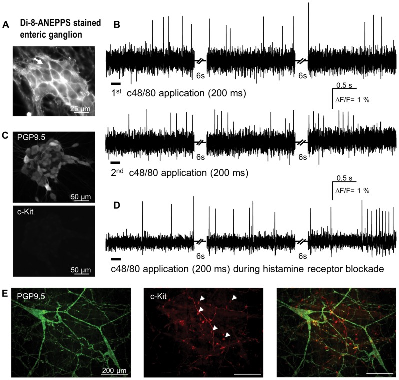
Figure 3. Compound 48/80 (c48/80) evoked spike discharge in primary cultured enteric neurons. A.
Di-8-ANEPPS stained ganglion; the dye incorporates into the outer membrane revealing the outline of individual neuronal cell bodies. B illustrates action potential discharge in response to two consecutive c48/80 applications (marked by bars below the traces) from the neuron marked by white arrow in A. The traces show responses during three recording periods with non-recording periods of 6 s in between. In this ganglion 17 of 19 neurons responded to c48/80. C PGP9.5-positive cultured enteric neurons. Lack of c-kit immunoreactivity demonstrated lack of mast cells in the culture. D demonstrates that c48/80 application still evokes spikes in cultures treated with a combination of the H1 and H2 blockers pyrilamine (1 µM) and ranitidine (10 µM), respectively. E shows green PGP9.5-positive neurons in a whole mount preparation of the guinea pig submucous plexus (left panel) and red c-kit-positive round and smooth mast cells (some marked by white triangles in center panel), which are morphologically distinct from the spindle shaped interstitial cells of Cajal. The right panel shows the merged image of the two stainings.

